Example case 39 of 40
The history began before admission
Recognise a subacute encephalitic syndrome when correction of a common abnormality leaves unexplained memory change and focal seizures.
Click or tap each illustration to enlarge it. The commentary and text transcript follow below.
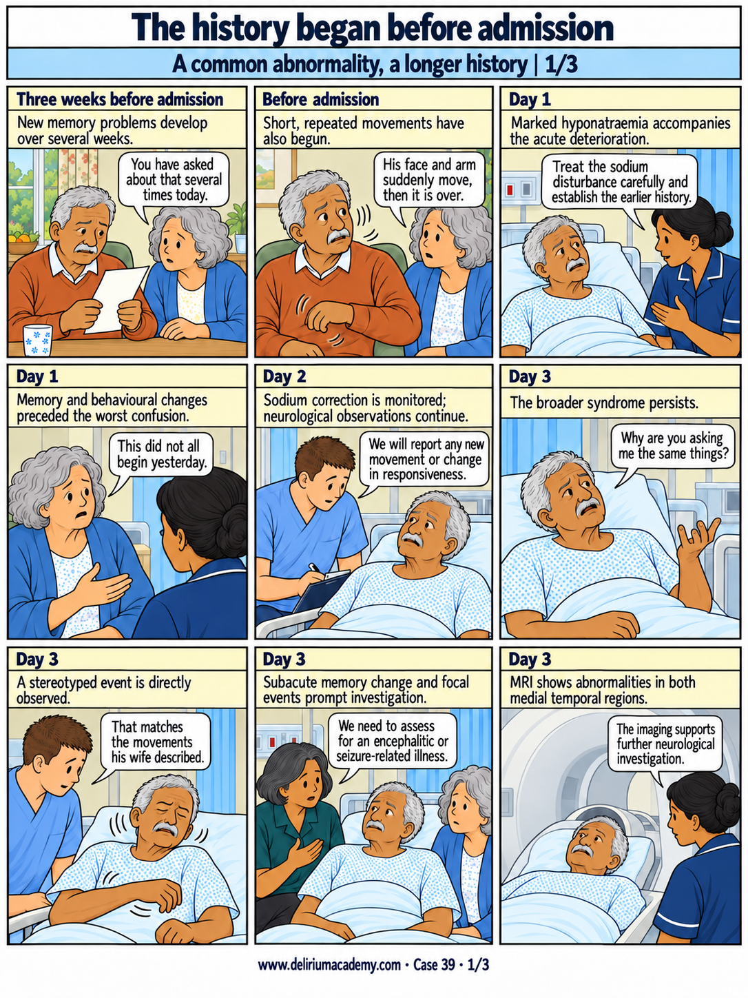
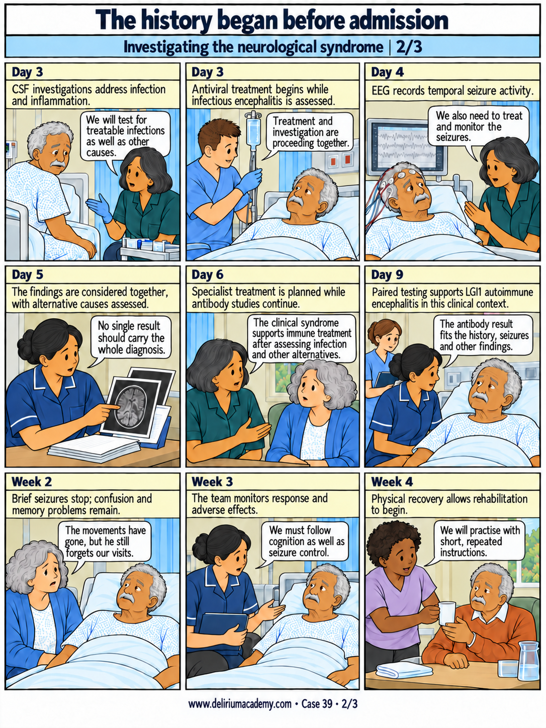
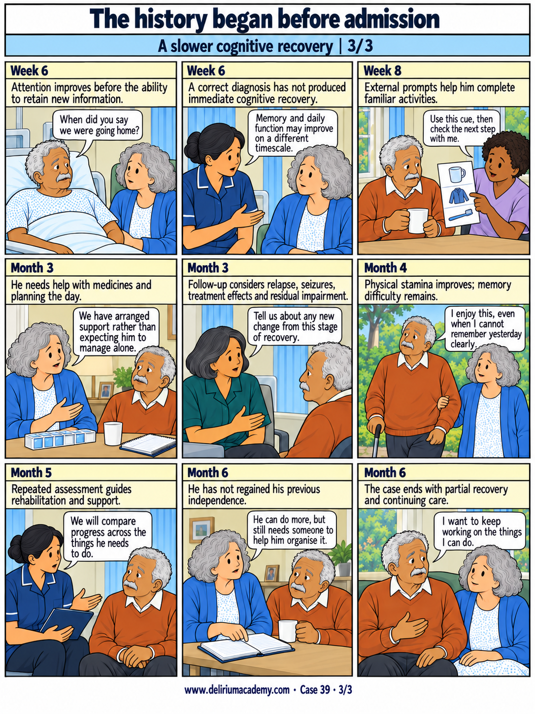
Read the text transcript
Page 1
Panel 1: Three weeks before admission
New memory problems develop over several weeks.
Celeste: You have asked about that several times today.
Panel 2: Before admission
Short, repeated movements have also begun.
Celeste: His face and arm suddenly move, then it is over.
Panel 3: Day 1
Marked hyponatraemia accompanies the acute deterioration.
Ward doctor Amira: Treat the sodium disturbance carefully and establish the earlier history.
Panel 4: Day 1
Memory and behavioural changes preceded the worst confusion.
Celeste: This did not all begin yesterday.
Panel 5: Day 2
Sodium correction is monitored; neurological observations continue.
Nurse Tom: We will report any new movement or change in responsiveness.
Panel 6: Day 3
The broader syndrome persists.
Harish: Why are you asking me the same things?
Panel 7: Day 3
A stereotyped event is directly observed.
Nurse Tom: That matches the movements his wife described.
Panel 8: Day 3
Subacute memory change and focal events prompt investigation.
Neurologist Mira: We need to assess for an encephalitic or seizure-related illness.
Panel 9: Day 3
MRI shows abnormalities in both medial temporal regions.
Ward doctor Amira: The imaging supports further neurological investigation.
Page 2
Panel 10: Day 3
CSF investigations address infection and inflammation.
Neurologist Mira: We will test for treatable infections as well as other causes.
Panel 11: Day 3
Antiviral treatment begins while infectious encephalitis is assessed.
Nurse Tom: Treatment and investigation are proceeding together.
Panel 12: Day 4
EEG records temporal seizure activity.
Neurologist Mira: We also need to treat and monitor the seizures.
Panel 13: Day 5
The findings are considered together, with alternative causes assessed.
Ward doctor Amira: No single result should carry the whole diagnosis.
Panel 14: Day 6
Specialist treatment is planned while antibody studies continue.
Neurologist Mira: The clinical syndrome supports immune treatment after assessing infection and other alternatives.
Panel 15: Day 9
Paired testing supports LGI1 autoimmune encephalitis in this clinical context.
Ward doctor Amira: The antibody result fits the history, seizures and other findings.
Panel 16: Week 2
Brief seizures stop; confusion and memory problems remain.
Celeste: The movements have gone, but he still forgets our visits.
Panel 17: Week 3
The team monitors response and adverse effects.
Ward doctor Amira: We must follow cognition as well as seizure control.
Panel 18: Week 4
Physical recovery allows rehabilitation to begin.
Occupational therapist Ruth: We will practise with short, repeated instructions.
Page 3
Panel 19: Week 6
Attention improves before the ability to retain new information.
Harish: When did you say we were going home?
Panel 20: Week 6
A correct diagnosis has not produced immediate cognitive recovery.
Ward doctor Amira: Memory and daily function may improve on a different timescale.
Panel 21: Week 8
External prompts help him complete familiar activities.
Occupational therapist Ruth: Use this cue, then check the next step with me.
Panel 22: Month 3
He needs help with medicines and planning the day.
Celeste: We have arranged support rather than expecting him to manage alone.
Panel 23: Month 3
Follow-up considers relapse, seizures, treatment effects and residual impairment.
Neurologist Mira: Tell us about any new change from this stage of recovery.
Panel 24: Month 4
Physical stamina improves; memory difficulty remains.
Harish: I enjoy this, even when I cannot remember yesterday clearly.
Panel 25: Month 5
Repeated assessment guides rehabilitation and support.
Ward doctor Amira: We will compare progress across the things he needs to do.
Panel 26: Month 6
He has not regained his previous independence.
Celeste: He can do more, but still needs someone to help him organise it.
Panel 27: Month 6
The case ends with partial recovery and continuing care.
Harish: I want to keep working on the things I can do.
Sources
- [S17] Jith, A., Mathew, K.A., Narayanan, D. and Jacob, S.M. (2021) ‘CASPR2 positive autoimmune encephalitis presenting as delirium in an alcohol dependent patient: a diagnostic dilemma’, Kerala Journal of Psychiatry, 34(1), pp. 54–56.
- [S18] Bandyopadhyay, S., Sahu, A., Chandra, S.K., Ghosh Dastidar, A. and Pratihar, S. (2026) ‘Autoimmune Encephalitis in Later Life: A Missed Opportunity in Routine Geriatric Care’, Cureus, 18(2), e104404.
- [S20] Yen, L., Leung, M., Kellaher, D.C., Kukoyi, O. and Xiong, G.L. (2011) ‘The Horse With Stripes: A Case of Anti-NMDA Receptor Encephalitis’, Primary Care Companion for CNS Disorders, 13(4), 11l01141.
- [G02] Graus F, Titulaer MJ, Balu R, et al. A clinical approach to diagnosis of autoimmune encephalitis. Lancet Neurology. 2016;15:391–404.
- [G01] National Institute for Health and Care Excellence. Delirium: prevention, diagnosis and management in hospital and long-term care. CG103.
Commentary
Harish's sodium disturbance is real and requires treatment, but the history began before the final deterioration. Several weeks of new memory difficulty, altered sleep and brief stereotyped movements suggest a broader neurological syndrome. Correction of sodium is therefore followed by continued assessment rather than a declaration that the cause has been fully addressed.
New focal seizures, bilateral medial temporal MRI abnormalities and the subacute cognitive course justify neurological investigation. Cerebrospinal fluid studies address infection and inflammation, and EEG characterises the events. Empirical antiviral treatment is started while an infectious encephalitis is being assessed. Paired antibody testing later supports LGI1 autoimmune encephalitis in a compatible clinical context. The diagnosis is not based on an isolated positive blood test [G02].
The supplied reports illustrate both confirmed antibody-associated disease and less certain proposed autoimmune explanations [S17, S18, S20]. The references differ in evidential strength. The composite presents a coherent diagnostic sequence with objective supporting findings, while retaining concurrent hyponatraemia as part of the illness rather than erasing it.
Treatment stops the brief seizures before it restores memory and daily functioning. The last page follows rehabilitation and review over months. Harish's outcome remains incomplete at six months, with difficulty retaining new information. The discussion should ask which features justified the additional investigations and how clinicians would distinguish ongoing inflammation, seizures, medication effects and residual injury during follow-up. A long delirium episode alone would not establish autoimmune encephalitis.
An anonymised composite case that does not describe one identifiable person and is not research evidence.